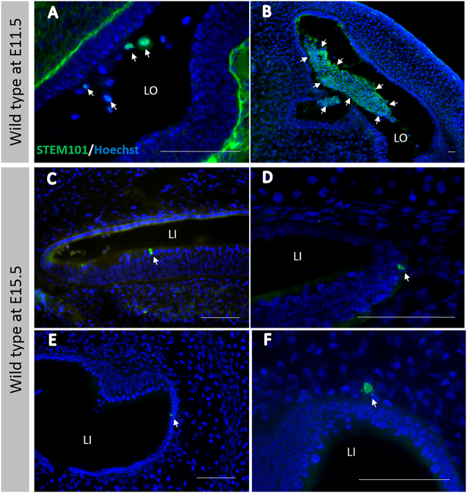
Figure 3.
(A) STEM101-positive cells in the lumen of the treated inner ears immediately after cell delivery into the otocyst. (B) Agglomerated STEM101-positive cells in the lumen of the treated inner ear (arrows) immediately (approximately 1 h) after cell delivery. (C) A STEM101-positive cell found in the cochlear epithelium (arrow) at the middle turn. (D) A STEM101-positive cell found in the cochlear lateral wall (arrow) at the apical turn. (E) A STEM101-positive cell in the semicircular canal epithelium (arrow). (F) A STEM101-positive cell in the subepithelium (arrow) at the treated semicircular canal. LO: Lumen of the otocyst. LI: Lumen of the inner ear. Green: STEM101; Blue: Hoechst. Scale bars indicate 100 μm.

