Figure 4.
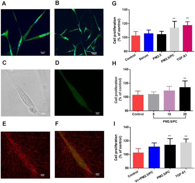
3D organotypic culture of human lung fibroblasts (HLFs). (A) Confocal image of fluorochrome-stained live cells and nuclei (magnification = ×40). (B) Confocal image of fluorochrome-stained live cells and the nucleus (magnification = ×20). (C) High magnification of the confocal image of a single HLF (magnification = ×40, zoom in factor = 2.5). (D) High magnification of the confocal image of a fluorochrome-stained single HLF (magnification = ×40, zoom in factor = 2.5). (E) High magnification of the confocal image of a fibrin-based scaffold (magnification = ×40, zoom in factor = 2.5). (F) Merged confocal image of a fibrin matrix covered by HLFs (magnification = ×40, zoom in factor = 2.5). Cell viability of 3D-cultured HLFs by stimulation of (G) control, serum, nanoscale PM2.5 (20 μg/ml), nanoscale PM2.5 (20 μg/ml)-protein corona (PC), and TGF-β1(2 ng/ml); (H) control and nanoscale PM2.5 (5, 10 and 20 μg/ml)-protein corona; (I) control and nanoscale PM2.5-protein corona/VC (vitamin C-0.1 mg/ml, nanoscale PM2.5–20 μg/ml); and nanoscale PM2.5 (20 μg/ml)-protein corona and TGF-β1 (2 ng/ml) (**P < 0.01, compared with the control).

