Fig. 2.
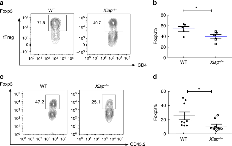
Foxp3 instability in activated Xiap−/− Treg cells. a, b Reduced Foxp3+ population in activated Xiap−/− tTreg cells. WT and Xiap−/− tTreg cells were stimulated with anti-CD3/CD28 in the presence of IL-2 for 4 days. Foxp3 expression of activated tTreg cells was determined by intracellular staining and analyzed by flow cytometry. The gated section represents the Foxp3+ population and the number indicates the percentage of each population (a). The Foxp3+ fractions in WT and Xiap−/− tTreg cells were quantitated (b), n = 5. c, d Diminished Foxp3high population in adoptively transferred Xiap−/− tTreg cells. CD45.2+ tTreg cells were recovered from CD45.1+ Rag1−/− mice into which CD45.2+ tTreg cells and CD45.1+ CD4+CD25− T cells had been transferred a month earlier. The isolated CD45.2+ CD4+ T cells were reactivated with TPA/A23187 and Foxp3 expression was determined by intracellular staining (c). The Foxp3+ fractions in the transferred WT and Xiap−/− tTreg cells re-isolated from Rag1−/− mice were quantitated (d), n = 9. *P < 0.05 for unpaired t-test

