Figure 1.
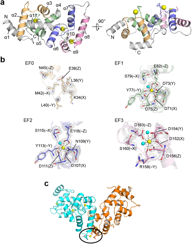
Structure of the Ca2+-bound form of calaxin. (a) Ribbon diagrams of the overall structure of calaxin in the Ca2+-bound open state. EF0, EF1, EF2 and EF3 are shown in light orange, pale green, slate and pink, respectively. Ca2+ ions are indicated by a yellow sphere model. (b) Close-up view of individual EF-hands binding a Ca2+ ion. Ca2+-binding residues are shown as a stick model. Ca2+ ions and water molecules are indicated by yellow and cyan spheres, respectively. The coordination bonds with Ca2+ ions are highlighted with dashed lines. The Fo − Fc omit maps of EF1, EF2 and EF3 are shown as gray meshes contoured at 3σ. (c) Calaxin dimer in the open state (cyan) and the closed state (orange) in an asymmetric unit. Ca2+ ions are represented as yellow spheres. Side chains coordinating Ca2+ between dimers are represented as sticks (surrounded by a black circle).

