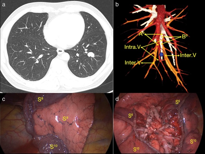
Figure 1.

Example of a case of laterobasale segmentectomy. (a) The tumor was located in the left lower lobe. (b) Three‐dimensional computed tomography bronchography and angiography identified targeted the segment bronchus, artery, and intrasegmental and intersegmental veins before surgery. The tumor (blue ball) was located in the left segment laterobasale. (c) Demarcation created by the modified inflation‐deflation method. The inflated segment was the targeted segment (S9) and S8 and S10 were the collapsed segments. (d) The remaining segments (S6, S8, S10) and stumps after segmentectomy. A9, laterobasale segment artery; B9, laterobasale segment bronchus; Intra.V, intrasegmental vein; Inter.V, intersegmental vein; S6, segment superius; S8, segment ventrobasale; S9, laterobasale segment; S10, segment dorsobasale.
