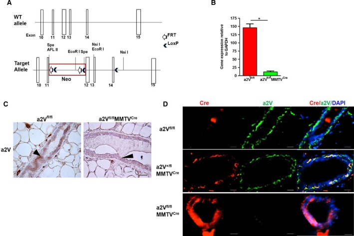
Figure 1.

Mammary epithelial cell‐specific deletion of a2V gene. (A) Schematic of the wild‐type and floxed ATP6V0a2 (a2V) gene. Exons 10–15 are shown with white boxes. The Lox/FRT‐Neo cassette was inserted upstream of exon 12 in an opposite direction relative to the a2V gene. A single LoxP site was inserted downstream of exon 14 in intron sequence. Some restriction enzyme sites are indicated. The presence of Cre and flox sites was confirmed by PCR (see Fig. S1A). (B) mRNA levels of a2 isoform in mammary epithelial cells isolated from breast tissues of a2Vfl/fl and a2Vfl/fl MMTVC re mice. n = 15, *P < 0.01. GAPDH was used as an endogenous control for normalization. The results are presented as mean ± SE. (C) Representative image showing a2V protein staining in mammary glands by immunohistochemistry (IHC) in breast tissues of a2Vfl/fl or a2Vfl/fl MMTVC re mice using anti‐a2V antibody. Brown color shows positive staining for a2V in mammary epithelial cells (black arrow head), and blue color shows nuclear staining by counterstain hematoxylin. n = 8, magnification 40×, scale bar 50 μm. (D) Representative image showing staining of Cre recombinase (red) and a2V (green) proteins in mammary glands of wild‐type (a2Vfl/fl), heterozygote (a2Vfl/+ MMTVC re), and a2V‐knockout (a2Vfl/fl MMTVC re) mice by immunofluorescence analysis (IFA). n = 8, magnification 20×, scale bar 100 μm.
