
Figure 4.
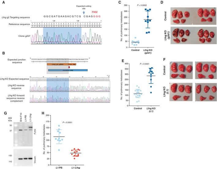
In vivo validation of the role of Lfng in metastasis. To validate the role of Lfng in metastasis, two independent Lfng targeting experiments were performed in B16‐F0 cells: one using a single gRNA to introduce a single base pair insertion (A, C and D) and another using two gRNAs to induce a 4.8‐kb deletion (B, E and F). (A) Sanger sequence trace of the targeted region in clone g2d1 carrying a homozygous 1‐bp insertion. (B) Image showing from top to bottom, the expected junction sequence after the deletion caused by the targeting of Lfng using two gRNAs, the expected reference sequence and the Sanger sequence traces observed and assembled with SeqMan Pro (Lasergene) against the expected reference sequence. The expected junction sequence separated by a single base insertion can be observed. Experimental metastasis assays using control and Lfng‐deficient cell lines (tail‐vein‐injected into wild‐type female mice (symbols representing individual mice with horizontal bar at the mean ± SD and statistics performed using a Mann–Whitney test; data shown are representative of two independent experiments)). Photographs are representative images of the lungs from mice injected with control and Lfng‐deficient cell lines. Plasmid rescue showing that introduction of the Lfng cDNA reverts the metastatic phenotype of L1 cells (L1‐Lfng; Lfng‐transfected cells, L1‐PB, vector‐only controls) (G and H). (G) A western blot with an anti‐Flag antibody shows restoration of Lfng expression (clone L1‐Lfng). An anti‐vinculin antibody was used as a loading control. These results are representative of three independent experiments. (H) Experimental metastasis assays using control L1‐PB cells and Lfng‐transfected cells. Please note experiments in e and h were performed with 5 × 10⁵ and 4 × 10⁵ cells, respectively, hence the different metastasis counts.