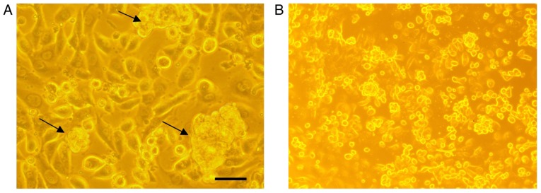
Figure 3.
Morphological examination of AntiGan-treated SW-480 cells. SW-480 cells were cultured in the (A) presence or (B) absence of 25 µl/ml AntiGan for 24 h. The morphological organization of the cells was examined by light microscopy. The black arrows indicate nuclear condensation. Magnification, ×40.

