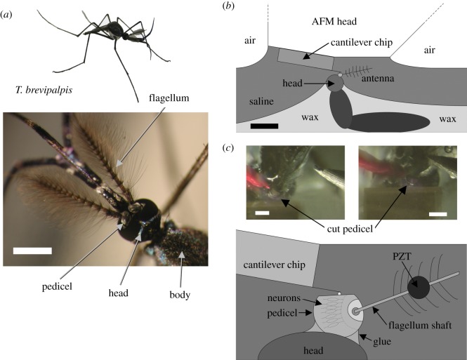
Figure 1.
Nanoscale in vivo and in situ force microscopy. (a) Male Toxorhynchites brevipalpis with close-up of head and plumose antennae. Scale bar, 1 mm. (b) Overall schematic of the preparation. Scale bar, 3 mm. (c) Close up schematic of the preparation. The PZT stimulus position is shown by the circle. Two photos of the preparation under the AFM are shown in the inset: left shows the preparation before the AFM's approach to the cut pedicel, right shows the AFM cantilever in position for measurements. Scale bars, 300 µm.

