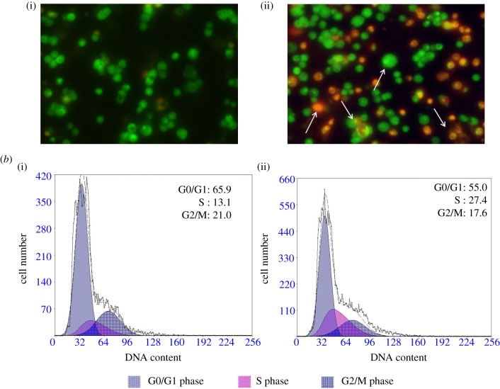
Figure 3.
(a) Morphological changes in LOVO cells treated with compound 7 g for 48 h: (i) LOVO control cells; (ii) LOVO cells treated with 5 µM for 48 h followed by morphological observation using AO/EB cell staining method. (b) Effect of LOVO on cell cycle progression of colon cancer cells: (i,ii) LOVO cells treated with 5 and 10 µM for 48 h followed by analysis of cell cycle distribution using propidium iodide cell staining method. Cell population in each cell cycle phase was numerically depicted. Data represent one of three independent experiments.

