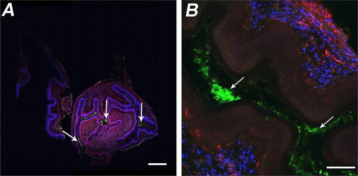
FIG 2 .

Confocal fluorescence micrographs of vaginal tissue excised from mice 4 h after i.vag. administration of FITC-BSA/ms. (A) FITC-BSA/ms (green) were located in the lumen (white arrows), but no CD4+ (red) or CD8+ (blue) cells were observed. Bar = 500 µm. (B) Higher magnification showing FITC-BSA/ms (green) located in the lumen (white arrows) and CD11b+ (blue) and CD11c+ (red) cells in the tissue. Bar = 100 µm.
