Figure 3.
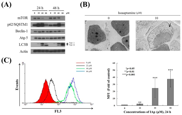
IAp induced autophagic hallmarks in T-47D cells. (A) Effect of IAp on the expression of autophagy-related proteins. Cells were treated with the indicated concentrations of IAp for 24 h and 48 h. Western blotting analysis was performed with mTOR, p62/SQSTM1, Beclin 1, Atg 5, and LC3B antibodies. Actin was the loading control. (B) Cells were treated with 44 μM of IAp for 24 h. Images of TEM were examined after treatment. (C) T-47D cells were treated with the indicated concentrations of IAp for 24 h. After treatment, cells were incubated with acridine orange for 30 min at 37 °C and analyzed using flow cytometry. Quantitative analysis of proton-pumping V-type ATPase activity showed a gradual increase of red fluorescent intensity upon IAp treatment when compared with the control group. Data are expressed as the mean ± SD of three experiments (* p < 0.05; ** p < 0.01; *** p < 0.001 compared with the control groups).

