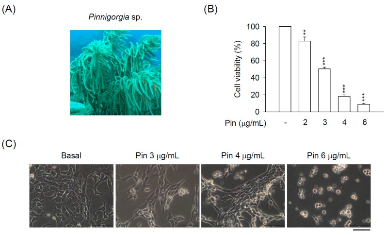
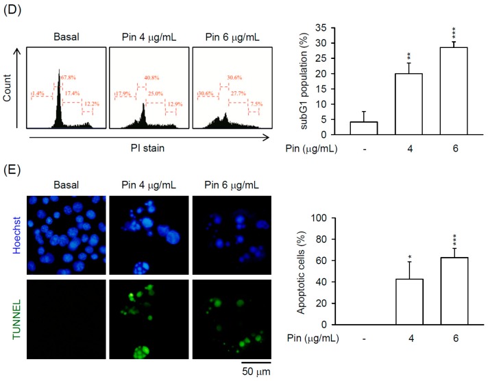
Figure 1.
Pin elicits apoptosis in hepatic stellate cells (HSCs). HSC-T6 cells were treated with Pin (0–6 µg/mL) for 24 h. (A) Gorgonian coral Pinnigorgia sp.; (B) Cytotoxicity assay was monitored spectrophotometrically at 450 nm; (C) Cell retraction, bubbling, and apoptotic bodies were observed using microscopy; (D) SubG1 population was examined by propidium iodide (PI) staining and flow cytometry; (E) The Pin-induced apoptosis of HSC-T6 cells was determined by terminal deoxynucleotidyl transferase dUTP nick end labeling (TUNEL) assay (green). Hoechst 33,342 (blue) was used to visualize the cell nucleus. All data are expressed as the mean ± S.E.M. (n = 3). * p < 0.05, ** p < 0.01, *** p < 0.001 compared with the basal.


