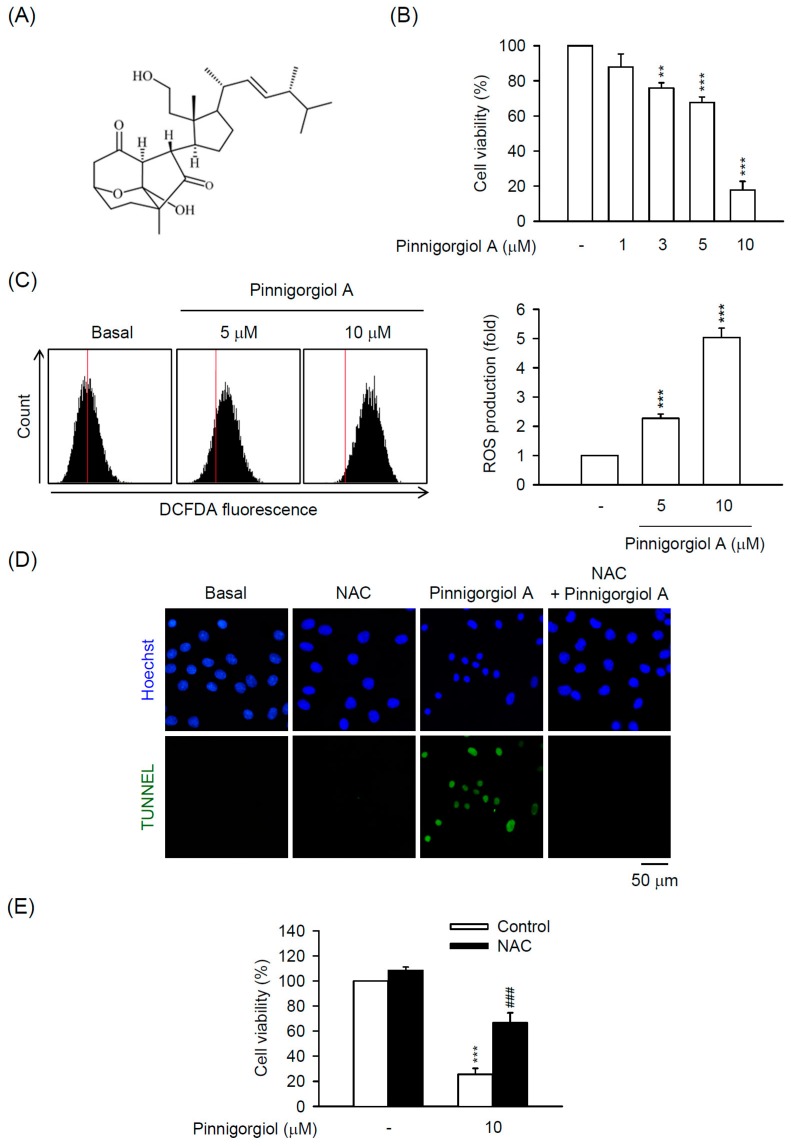
Figure 6.
Pinnigorgiol A exhibits ROS-dependent apoptosis in HSCs. (A) Chemical structure of pinnigorgiol A; (B) HSC-T6 cells were treated with pinnigorgiol A (1–10 µM) for 24 h. Cytotoxicity assay was monitored spectrophotometrically at 450 nm; (C) HSC-T6 cells were treated with pinnigorgiol A (5 or 10 µM) for 6 h. Intracellular ROS was determined by DCFDA assay and flow cytometry; (D) HSC-T6 cells were treated with pinnigorgiol A (10 µM) for 12 h. Apoptotic effect of pinnigorgiol A was determined by TUNEL assay (green). Hoechst 33,342 (blue) was used to visualize the cell nucleus; (E) HSC-T6 cells were pretreated with NAC (2.5 mM) for 1 h and then incubated with pinnigorgiol A (10 µM) for 24 h. Cytotoxicity assay was monitored spectrophotometrically at 450 nm. All data are expressed as the mean ± S.E.M. (n = 3). ** p < 0.01; *** p < 0.001 compared with the basal. ### p < 0.001 compared with the pinnigorgiol A alone.

