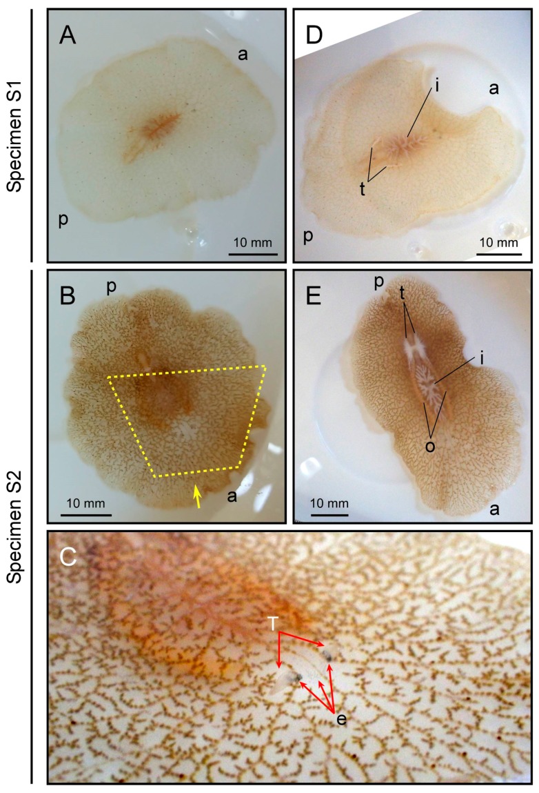
Figure 2.
External morphology of polyclad flatworms. Upper and lower panels represent specimens S1 and S2, respectively. Panel (A,B) dorsal views; Panel (C) anterior region indicated by the yellow dotted line from the direction of the yellow arrow in panel (B); Panel (D,E) ventral views. a and p represent anterior and posterior ends of the body, respectively. t, testis; o, ovary; T, tentacle; e, eye spots; i, intestine.

