Figure 5.
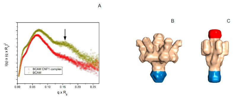
Small angle X-ray analysis of Lu/BCAM complex formation with CNF1. (A) Normalized Kratky plots of Lu/BCAM and Lu/BCAM-CNF1 complex. The Kratky plot of Lu/BCAM (dark yellow trace) shows that Lu/BCAM is an aspherical particle with large flexible areas as indicated by an arrow. In contrast, the normalized Kratky plot of the Lu/BCAM-CNF1 complex (red trace) reveals that the flexibility is largely reduced; (B) SAXS envelope reconstructed from the scattering curve with DAMMIF of Lu/BCAM assuming a six-fold symmetry as calculated from the size of the particle. The presumable Fc region of recombinant Lu/BCAM fusion protein is indicated in blue. The protruding arm-like structures correspond in size to the five Ig domains of the extracellular domain of Lu/BCAM, respectively; (C) The reconstructed envelope of Lu/BCAM-CNF1 complex shows additional density at the top end of the particle and indicates largely reduced flexibility of the arm-like structures, which assemble in the center of the molecule. This is strongly supported by the normalized Kratky plot depicted in (A).

