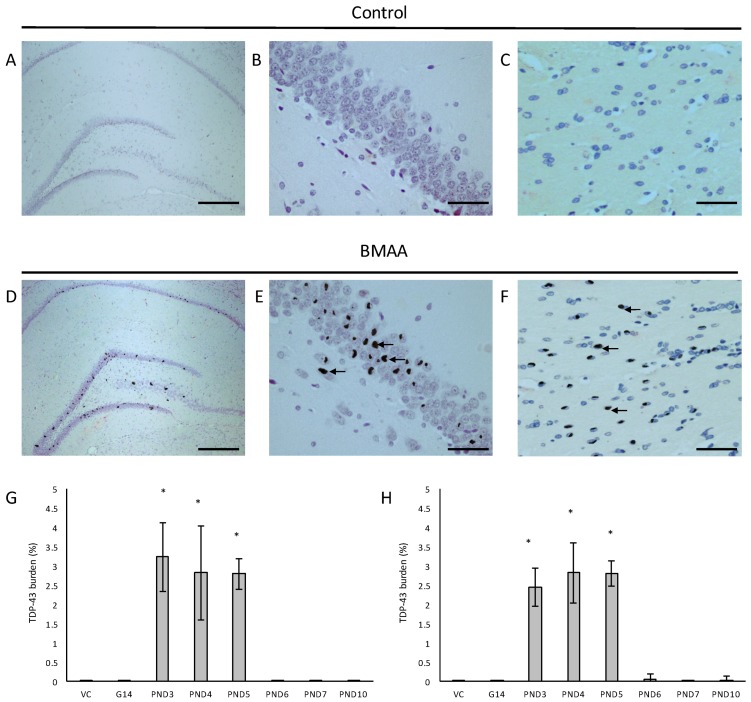
Figure 8.
Neurons with pathological TDP-43 positive cytoplasmic inclusions (indicated by arrows) in the hippocampal formation (D–F) of male (D) and female (E,F) rats exposed to BMAA on PND3 and not in gender-matched rats exposed to the vehicle control on PND3. The percentage pathological TDP-43 burden, defined as the percentage neurons in the hippocampus that harbor hyperphosphorylated TDP-43 positive inclusions, is given for all male (n = 5) (G) and female (n = 5) (H) exposure groups. Scale bars correspond to 400 μm (A,D) and 30 μm (B,C,E,F). * indicates significant difference to the control (p < 0.05).

