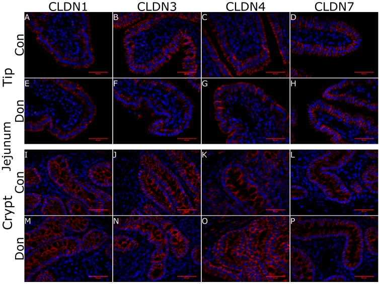
Figure 5.
Claudin surface localization in piglet jejunal villi and crypts in DON-fed and control fed piglets. Jejunal tissue was obtained 24 days after DON-exposure to half of the piglets. CLDN1 was localized to the full length of the pericellular junction within the crypts where as it was found more heavily localized to the apical aspect of the pericellular junction at the villus tip (A,E,I,M). CLDN3 stained the length of the pericellular junction at the villus tip and within the crypts but was more abundant in the latter (B,F,J,N). CLDN4 stained the villous surface but was found intracellularly localized in the epithelium of the crypts (C,G,K,O). CLDN7 stained along the length of the pericellular junction at both the villus tip and within the crypts (D,H,L,P). Secondary antibody: Alexa555-conjugated goat α rabbit IgG (red) in incubation buffer for 4 h at room temperature. Nuclear stain: DAPI (blue). Scale bar represents 50 μm.

