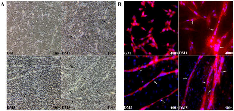
Figure 1.
(A) Microscopic images of chicken primary myoblasts cultured in growth medium (GM) (100% confluence) or in differentiation medium (DM) for 24 h (DM1), 72 h (DM3) and 120 h (DM5). Black arrows represent myotubes; and (B) cells were fixed and immunostained for desmin in GM and DM. Nuclei were stained with DAPI. White arrows represent fused myotubes. Cells at different developmental stages were collected for use in the following procedures. The red and blue color represented cytoplasm and cell nucleus, respectively.

