Fig. 7.

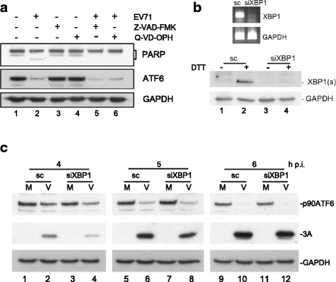
Effects of host proteinases and caspases on the virus-induced decrease in ATF6. a MCF7 cells were infected with EV71 and then treated with Z-VAD-FMK (20 μM) and Q-VD-OPH (20 μM) at 8 and 0 h p.i., respectively. Cell lysates were harvested at 10 h p.i. and evaluated by western blotting. b (Upper panel) Semiquantitative PCR analysis of XBP1 mRNA in RD cells transfected with siXBP1 or scrambled siRNA. (Bottom panel) RD cells were transfected with siXBP1 and then treated with 2.5 mM DTT, an ER stress inducer, for 6 h. The level of XBP1(s) was detected by western blotting. c RD cells were transfected with siXBP1 or scrambled siRNA for 72 h, infected with EV71 at an MOI of 10, and harvested at the indicated times after infection. The ATF6 levels in the virus-infected XBP1-silenced cells were detected by western blotting
