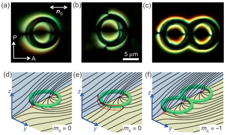
Figure 16.
(a,b) Cross-polarized images of a toroidal particle with perpendicular surface anchoring of the liquid-crystal molecules immersed in a planar nematic cell. Two point defects can be seen in panel (a). In panel (b) one point defect has opened into a small loop. (c) A handlebody with genus is accompanied by three point defects. (d–f) Schematic drawing of the director field for the three images in (a–c). The scale bar is 5 µm. Adapted from Ref. [70].

