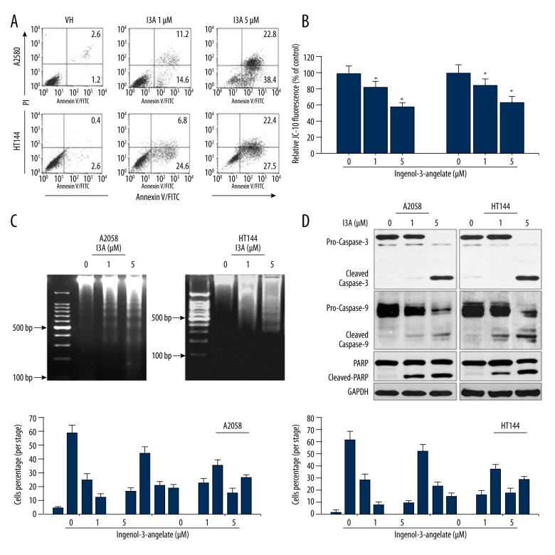
Figure 2.
I3A induces apoptotic cells death and cell cycle arrest. (A) Flow cytometry analysis of A2058 and HT144 cells treated with or without I3A after annexin V/PI-FITC staining. (B) ELISA microplate measurement of mitochondrial membrane potential using JC-10 staining of A2058 and HT144 cells treated with or without I3A. Data represent percent of control from experimental triplicate. (C) Agarose gel electrophoresis of genomic DNA isolated from A2058 and HT144 cells treated with or without I3A. Percent colony formation is represented as compared to control. (D) Western blot analysis of apoptosis-related proteins from A2058 and HT144 cells treated with or without I3A. (E) Flow cytometry analysis of cell cycle from A2058 and HT144 cells treated with or without I3A after PI staining. Each quantitative experiment was performed in triplicate. I3A – ingenol-3-angelate; * P<0.05 vs. control.

