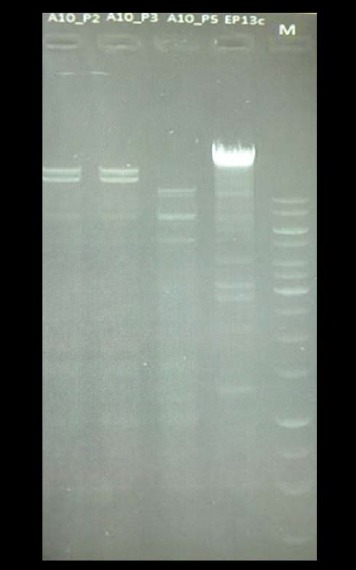
Fig. 3.

Restriction with PvuII of DNA from three different E. coli phage isolates from one sampling site, drain on floor (Table 2). From left to right: slot 1, phage number 2; slot 2, phage number 3; slot 3, phage number 5; slot 4, control phage; slot 5, molecular weight marker. Phage numbers 2 and 3 gave the same restriction pattern, and phage number 5 differs from those two
