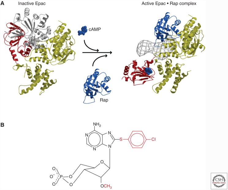
Figure 3.
(A) Crystal structure of Epac 2 in the closed (left) and open (right) conformation. The gray area (right) represents the first cyclic adenosine monophosphate (cAMP) domain and Dishevelled, EGL-10, and pleckstrin (DEP) domain visualized by cryoelectron microscopy (Rehmann et al. 2006, 2008). (B) Structure of 8CPT-2′OMe-cAMP (007).

