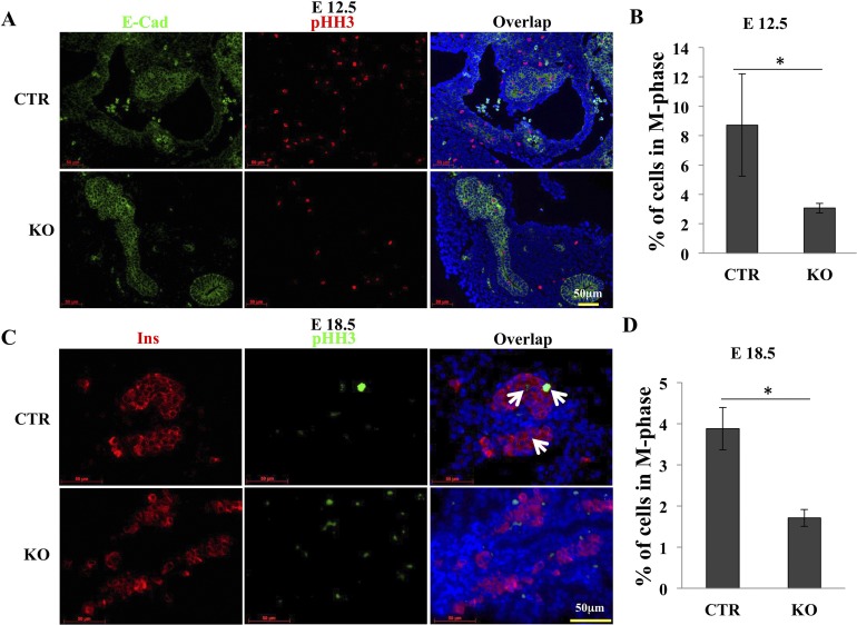
Figure 4.
Deletion of GRP94 reduces proliferation of Pdx1+ cells at E12.5. (A) Representative immunohistochemical staining of pancreases from CTR (n = 4) and KO (n = 5) embryos at E12.5 using anti–E-cadherin (green) and anti-pHH3 (red) antibodies. Nuclei are stained blue. Scale bar = 50 μm. (B) Percentage of pHH3+ cells in Pdx1+ cells. (C) Representative immunohistochemical staining of pancreases from CTR (n = 4) and KO (n = 4) embryos at E18.5 using anti-insulin (Ins; red) and anti-pHH3 (green) antibodies. Nuclei are stained blue. Arrows point to pHH3+Ins+ cells. Scale bar = 50 μm. (D) Percentage of pHH3+ cells in Ins+ cells.

