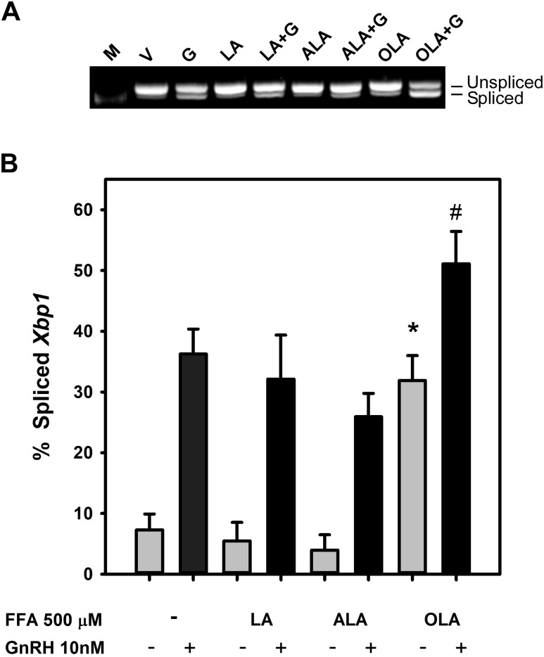
Figure 1.
FFA induction of Xbp1 mRNA splicing in LβT2 gonadotropes. LβT2 cells were serum-starved for 12 to 16 hours prior to treatment with vehicle (V) or 500 µM LA, ALA, or OLA for 3 hours with or without 10 nM GnRH (G) for the final 30 minutes. Total RNA was isolated, and Xbp1 was analyzed by RT-PCR using primers that cross the 26-bp intron that is spliced in response to activation of the UPR by the ER-resident RNAse ERN1. Bands are visualized in comparison to 500-bp DNA ladder (M). (A) Agarose gel electrophoresis of RT-PCR products showing the presence of unspliced (top band) and spliced (bottom band) Xbp1 mRNA. (B) Summary of densitometric quantification of digitally acquired agarose gel images from three independent experiments showing Xbp1 splicing induced by OLA or GnRH, but not LA, or ALA. Bars represent the percentage of spliced form of Xbp1 relative to total detected. Error bars denote standard error of the mean of at least three independent experiments. *Significant difference from vehicle control; #significant difference from GnRH-treated control by ANOVA and Tukey post hoc multiple-comparison test. All GnRH-treated groups are significantly increased from their respective untreated control.

