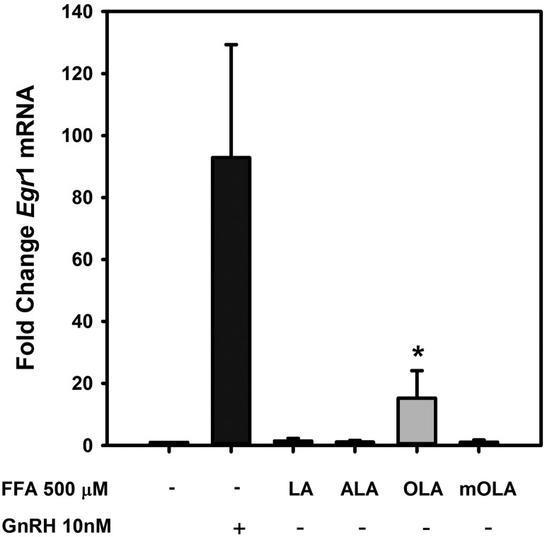
Figure 4.
FFA induction of Egr1 expression in LβT2 gonadotropes. LβT2 cells were serum-starved for 12 to 16 hours and then treated for 3 hours with vehicle or 500 µM LA, ALA, OLA, or mOLA with or without treatment with 10 nM GnRH for the final 30 minutes of incubation. At the end of incubation, cells were harvested, and total RNA was prepared from cell extracts. Egr1 mRNA expression was analyzed by quantitative real-time PCR using Gapdh as an internal reference control. Bars summarize the fold change of Egr1 mRNA after treatment relative to untreated vehicle control from three independent experiments. The corresponding GnRH treatment groups are provided in Supplemental Fig. 1 (24.2MB, tif) . The results show that both GnRH and OLA treatment increases Egr1 mRNA. Cotreatment with OLA and GnRH showed no additive or synergistic effects, and treatment with LA, ALA, and mOLA either alone or with GnRH treatment had no effect (Supplemental Fig. 1 (24.2MB, tif) ). Error bars denote standard error of the mean of at least three independent experiments. *Significant difference from untreated control as determined by ANOVA and post hoc testing with Dunnett comparison with control test.

