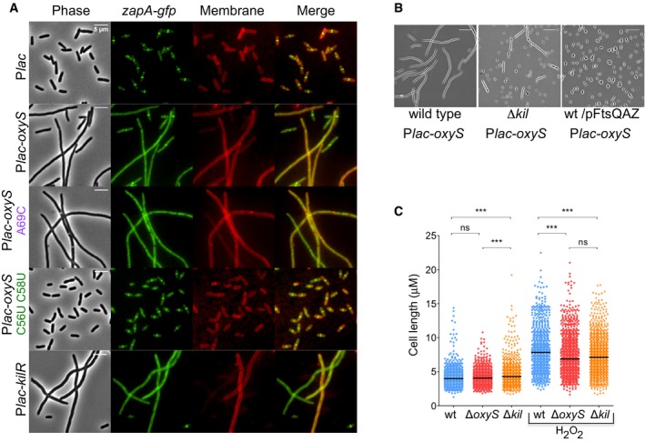
Figure 5. Fluorescence microscopy images.

- OxyS expression impairs cell division. Cultures of Escherichia coli carrying ZapA‐gfp fusion as a single copy in the native position in the chromosome (ZapA‐gfp:cat) and Plac‐oxyS plasmids were treated with 1 mM IPTG at dilution. Samples were taken at 3 h post‐dilution. All samples were spotted on PBS agar pad for imaging. DNA stained blue with DAPI. Scale bar, 5 μm.
- OxyS‐mediated impaired cell division is prevented in ΔkilR mutant and by overexpression of FtsQAZ. Cells were grown as described above for 3 h at 37°C in the presence of 1 mM IPTG. The operon FtsQAZ is expressed from its own promoter. Scale bar, 5 μm.
- OxyS induced by H2O2 impairs cell division. Scatter plots of cell length distribution of wild‐type ΔoxyS and ΔkilR grown without or with H2O2 treatment. The cultures at OD600 = 0.1 were exposed to 1 mM H2O2 or remained untreated. Cell lengths were measured 60 min thereafter. The black line in each plot represents the median of three biological experiments. In each experiment, more than 750 cells were analyzed (GraphPad Prism software; unpaired t‐test, ***P‐value = 0.0001).
