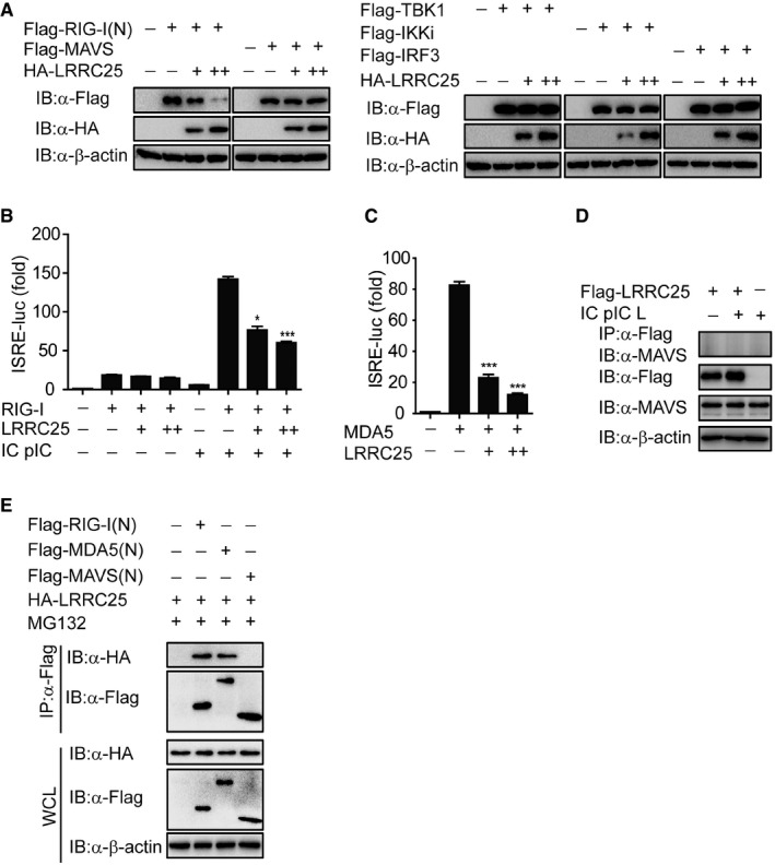
Figure EV3. LRRC25 cannot interact with MAVS, related to Fig 3 .

- The expressions of RIG‐I (N), MAVS, TBK1, IKKi, IRF3, and LRRC25 in Fig 3A were analyzed by IB analysis.
- HEK293T cells were transfected with an empty plasmid or increasing amounts of plasmid for LRRC25, plus an ISRE‐luc reporter and plasmids for RIG‐I. After 12 h, cells were left untreated or treated with IC poly(I:C) (5 μg/ml) for 24 h. Cell lysates were analyzed for ISRE‐luc activity.
- HEK293T cells were transfected with an empty plasmid (no wedge) or increasing amounts (wedge) of plasmid for LRRC25, plus an ISRE‐luc reporter and plasmid for MDA5. 24 h post‐transfection, cell lysates were analyzed for ISRE‐luc activity.
- HEK293T cells were transfected with Flag‐LRRC25. After 12 h, the cells were left untreated or treated with IC poly(I:C) LMW (5 μg/ml) for 24 h. Cell lysates were immunoprecipitated using anti‐Flag, followed by immunoblots using the indicated antibodies.
- HEK293T cells were transfected with Flag‐RIG‐I (N), Flag‐MDA5 (N), Flag‐MAVS (N), and HA‐LRRC25 for 24 h. Before harvesting, the cells were treated with DMSO or MG132 (5 μM) for 4 h. Cell lysates were immunoprecipitated using anti‐Flag, followed by immunoblot using the indicated antibodies.
