-
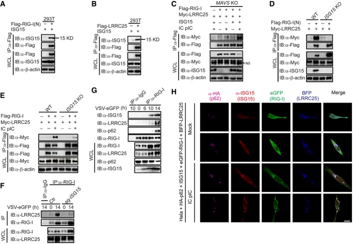
A, B
HEK293T cells were transfected with ISG15, together with Flag‐RIG‐I (N) (A) or Flag‐LRRC25 (B). 24 h post‐transfection, cell lysates were immunoprecipitated using anti‐Flag, followed by immunoblot using the indicated antibodies.
-
C
MAVS KO HEK293T cells were transfected with Flag‐RIG‐I and Myc‐LRRC25, together with an empty vector or ISG15. After 12 h, the cells were left untreated or treated with IC poly(I:C) (5 μg/ml) for 24 h. Cell lysates were immunoprecipitated using anti‐Flag, followed by immunoblots using the indicated antibodies. NS indicates non‐specific bands.
-
D
WT and ISG15 KO COS7 cells were transfected with Flag‐RIG‐I (N) and Myc‐LRRC25. 24 h post‐transfection, cell lysates were immunoprecipitated using anti‐Flag, followed by immunoblot using the indicated antibodies.
-
E
WT and ISG15 KO COS7 cells were transfected with Flag‐RIG‐I and Myc‐LRRC25. After 12 h, the cells were left untreated or treated with IC poly(I:C) (5 μg/ml) for 24 h. Cell lysates were immunoprecipitated using anti‐Flag, followed by immunoblots using the indicated antibodies.
-
F
Control and ISG15 KO THP‐1 cells were infected with VSV‐eGFP (MOI = 0.1) for 14 h. Cell lysates were immunoprecipitated using anti‐RIG‐I, followed by immunoblots using anti‐LRRC25.
-
G
THP‐1 cells were infected with VSV‐eGFP (MOI = 0.2) for indicated time points, and cell lysates were immunoprecipitated using anti‐RIG‐I, followed by immunoblots using indicated antibodies.
-
H
Confocal microscopic analysis of HeLa cells co‐transfected with HA‐p62, ISG15, eGFP‐RIG‐I, and BFP‐LRRC25, followed IC poly(I:C) (5 μg/ml) treatment for 24 h. Scale bar, 20 μm.
Data information: In (A–H), data are representative of three independent experiments.

