Figure 5.
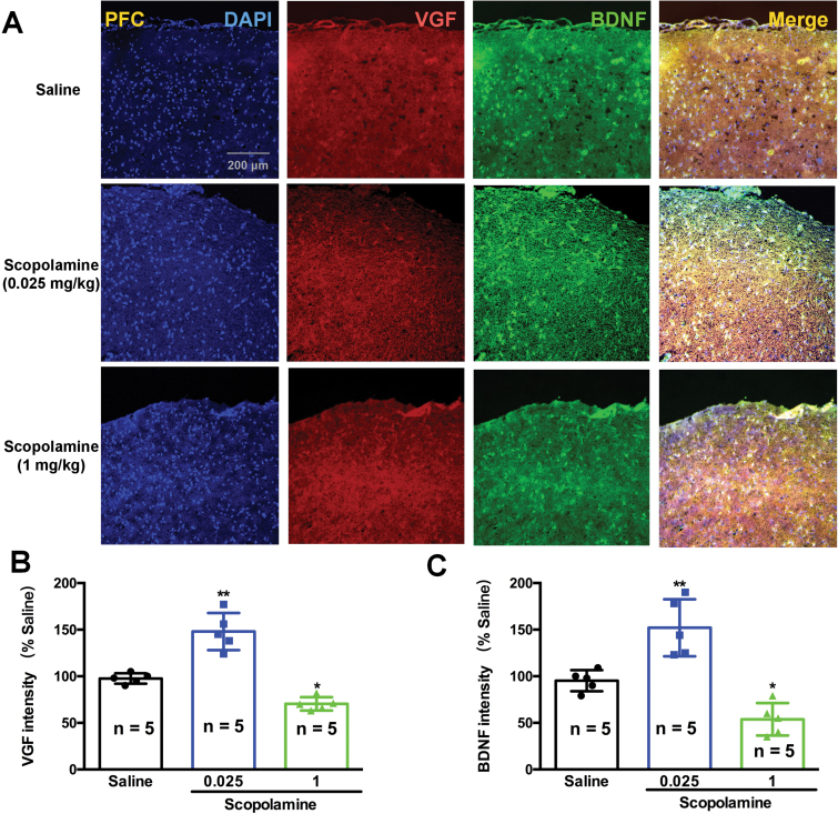
Effects of various doses of scopolamine on VGF and brain derived neurotrophic factor (BDNF) immunoreactivity and colocalization in the prefrontal cortex of mice. (A) VGF and BDNF protein expression was examined using immunofluorescence of frozen mice prefrontal cortex sections. Anti-VGF was labeled with an Alex Fluor 594 conjugated secondary antibody (red). Anti-BDNF antibody was labeled with an Alex Fluor 488 conjugated secondary antibody (green). The densities of both VGF and BDNF were significantly increased by a low dose of scopolamine (0.025 mg/kg) and were significantly decreased by a high dose of scopolamine (1 mg/kg). The cellular localization of VGF and BDNF after scopolamine treatment was characterized by colocalization studies with markers of neurons (DAPI). n=5/group. *P<.05, **P<.01 compared with the control (saline plus saline)group.

