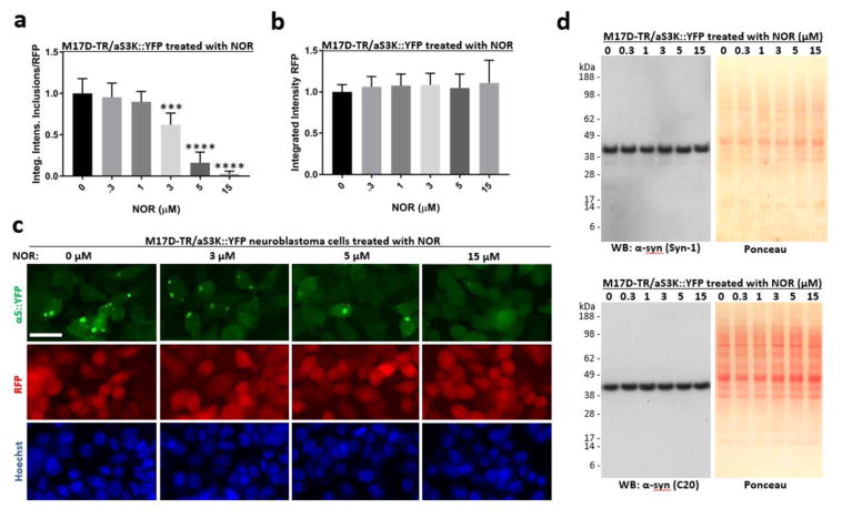
Figure 2. Nortriptyline reduces inclusion formation in M17D neuroblastoma cells that express inclusion-prone αS3K::YFP.
A) M17D cells that express an αS-3K::YFP fusion protein (dox-inducible) and RFP (constitutive) were treated with NOR at 0 (= DMSO alone at 0.1% f.c.), 0.3, 1, 3, 5 and 15 μM. αS-3K::YFP expression was induced at the same time point and IncuCyte-based analysis of punctate YFP signals relative to total RFP was performed 24 h later. YFP inclusion integrated intensities and RFP total integrated intensities were measured and the ratio was calculated. Four independent experiments (N = 4, n = 12), the data are presented as mean ± SD. ***p<0.001, ****p<0.0001 versus 0 μM NOR (whose mean was set to 1 in each experiment), repeated-measures one-way ANOVA followed by Dunnett’s multiple comparisons post hoc test. B) Same as A, but only the total integrated intensities of co-expressed RFP are plotted. No significant changes were observed. C) Representative images (YFP, RFP and Hoechst staining) for the indicated NOR concentrations; scale bar = 20μm. D) Western blots using α-syn-specific mAb Syn-1 and pAb C20, plus Ponceau-stained membranes.

