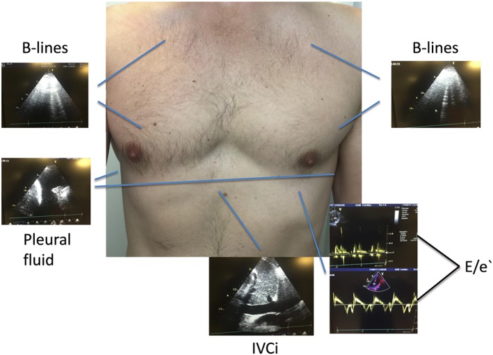
Figure 1.

Cardiothoracic ultrasound protocol showing B‐lines on lung ultrasound as a sign of congestion, pleural fluid, a typical mitral inflow, and tissue Doppler signals used to calculate the E/e′ ratio, as well as a subcostal view of the IVC. E/e′, E/e′ ratio medially; IVC, inferior vena cava.
