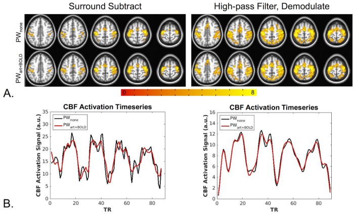
Fig 7. Finger-tapping task PW results.
(A) For the SS results (left), bilateral activation was observed in the motor cortex for the PWss,none and PWDNss,art+BOLD data. An increased activation area was seen for the PWDNss,art+BOLD data compared to the PWss,none data. The HD data (right) showed increased activation compared to the SS data, however no differences were seen between the denoised and non-denoised data the. All maps were thresholded at p<0.005 and cluster corrected with a minimum cluster size of 131 voxels (α<0.05). (B) Average SS PW signal from one representative subject (left) and average HD PW signal from the same subject (right). All PW signal was extracted from a mask of voxels active for all PW datasets. The denoised SS time-series appear less noisy with less variance compared to the non-denoised time-series. This effect is less apparent for the HD data.

