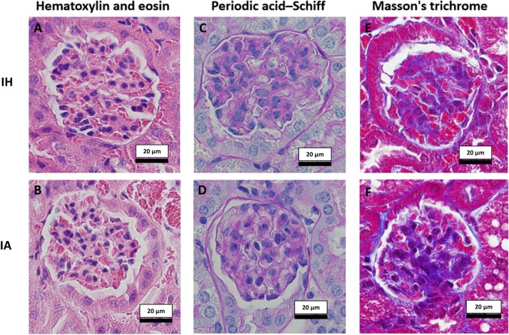
Fig 2. Examples of stained kidney sections used for subjective histological evaluation.
Two randomly chosen slides (4 sections per slide) from each group were evaluated by an independent histopathologist after being stained with hematoxylin and eosin (2A (IH), 2B (IA)) for general glomerular and tubular morphological assessment, cellularity of the glomerulus, and for cellular infiltrates in the cortex and medulla. Glomeruli stained with periodic acid–Schiff stain for glomerular basement membrane and mesangium assessment, and for assessment of glomerular capillary loops and tubular epithelium are shown in Fig 2C (IH) and 2D (IA). Assessments of glomerular and tubular collagen and fibrous tissue were made using Masson's trichrome stain; 2E (IH), 2F (IA). Images taken at 400x with standardized light exposure. IH: Intermittent hypoxia, IA: Intermittent air.

