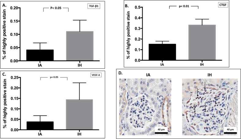
Fig 4. Glomerular expression of growth factors by immunohistochemistry.
Semi-quantitative analysis of glomerular expression of TGF-β1 (4A), CTGF (4B) and VEGF-A (4C) proteins by immunohistochemistry. Fig 4D indicates an examples of glomerular protein (CTGF) expression by microscopic viewing. Images taken at 400x magnification; nucleus indicated as blue, while brown staining indicates antigen-antibody reaction inside glomerular cells. Arrows indicate glomerular protein localization. IA: Intermittent air, IH: Intermittent hypoxia; unpaired t-test, (n = 5).

