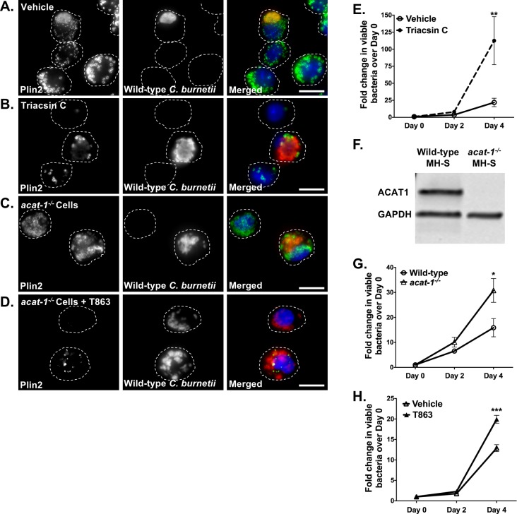
Fig 3. Blocking LD formation increases C. burnetii growth.
Wild-type C. burnetii growth in infected MH-S cells treated with different inhibitors was measured at 2 and 4 days post-infection by FFU assay. A-D) Representative images for wild-type MH-S macrophages treated with inhibitors, fixed, stained for PLIN2 (LDs; green) and C. burnetii (red) and imaged day 4 post-treatment at 100X. Scale bar = 10 µm. E) Growth while inhibiting LD formation with triacsin C (10 µM) in wild-type MH-S macrophages. Error bars represent the mean of 4 independent experiments +/- SEM. ** = p <0.01 compared to vehicle-treated cells as determined by two-way ANOVA with Bonferroni post-hoc test. F) ACAT1 protein expression in wild-type and acat-1-/- macrophages. Cell lysates were immunoblotted and ACAT1 protein levels were compared with GAPDH as loading control. G) C. burnetii growth in vehicle-treated wild-type and acat-1-/- MH-S macrophages and (H) T863-treated acat-1-/- MH-S macrophages. Error bars represent the mean of at least 3 independent experiments +/- SEM., * = p<0.05, *** = p <0.001 as determined by two-way ANOVA with Bonferroni post-hoc test.

