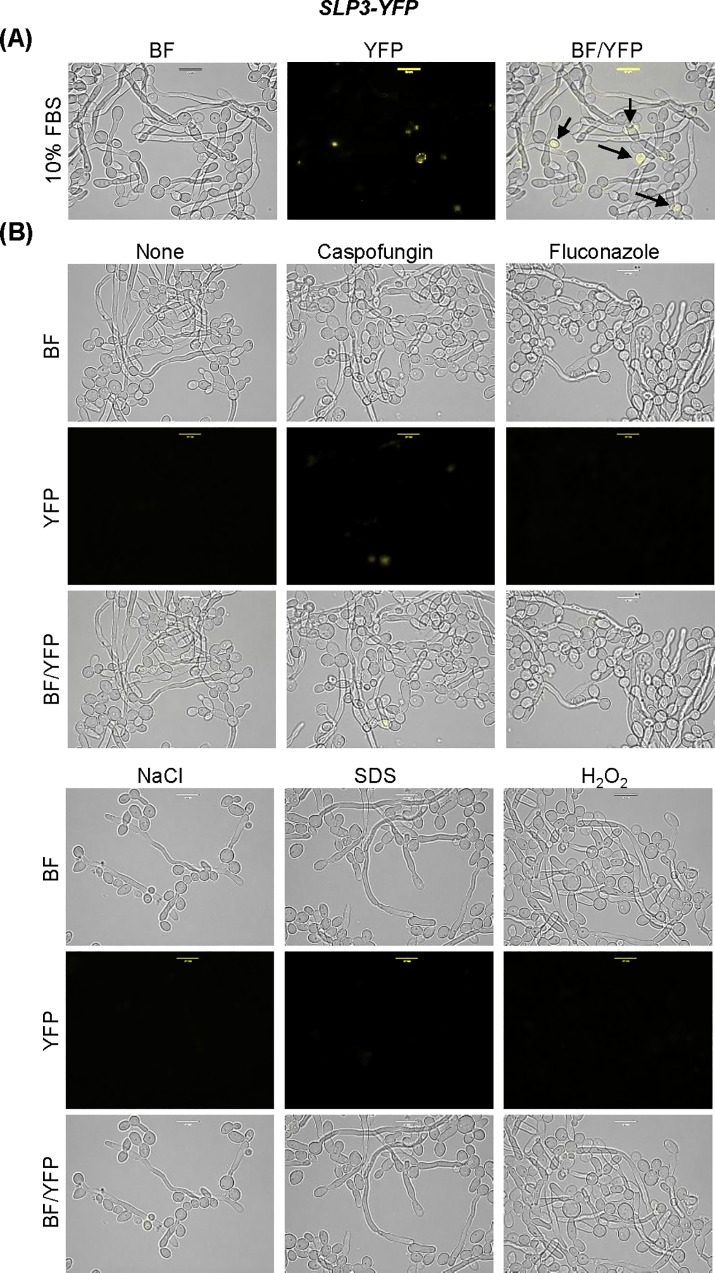
Fig 5. Localization of Slp3p in the yeast-to-hyphae transition.
(A) SLP3-YFP cells were grown in 10% FBS/YPD+uri medium for 16 hours and observed under bright-field and fluorescent microscopy. Arrows highlight fluorescent yeast-phase cells. (B) Overnight cultured SLP3-YFP hyphal cells were standardized in 10% FBS/YPD+uri media and incubated for 45 minutes with the given additives and viewed under bright-field and fluorescent microscopy. The concentration of additives used is as follows: 125 ng/mL caspofungin, 2.5 μg/mL fluconazole, 1.0 M NaCl, 0.17% H2O2, and 0.08% SDS. For each assay, three biological replicates were analyzed. Experiments were repeated at least three times, and data presented represents one representative experiment. Approximately 1.0 x 10⁴ cells of each strain were selected for viewing. Scale bars represent 10 μm.

