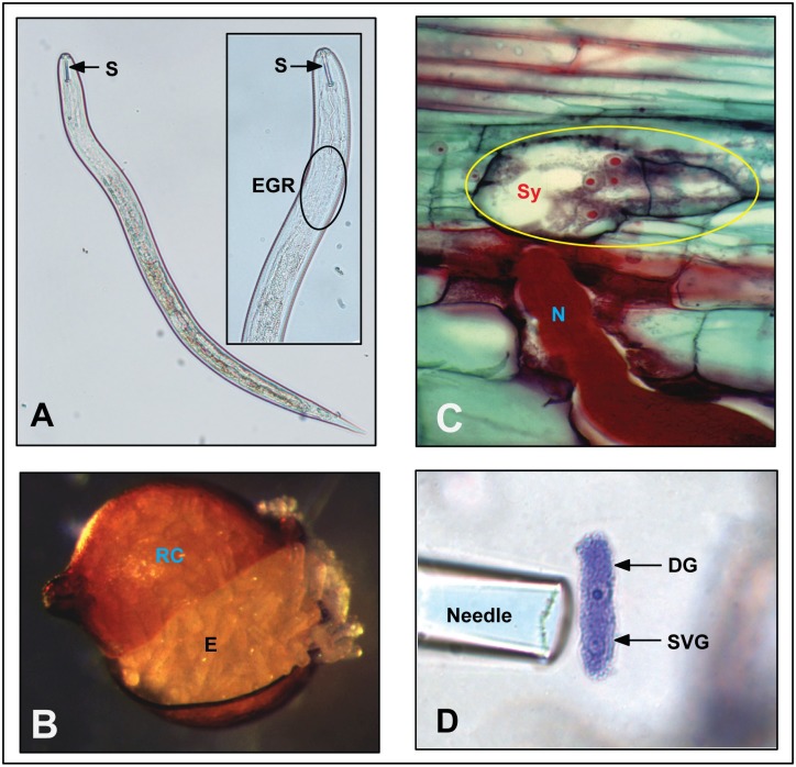
Fig 1.
A. Infective Heterodera glycines juvenile with a close-up of the anterior region showing the stylet (S) and the EGR. B. Ruptured RC displaying stored E. Image by E. C. McGawley, Nemapix. C. Cross section through a SY of a H. glycines N. Image by Burton Y. Endo, Nemapix. D. Microaspiration of isolated and stained DG and SVG gland cells of H. glycines showing prominently stained nuclei. Image by Thomas R. Maier, Iowa State University. DG, dorsal gland; E, eggs; EGR, esophageal gland region; N, nematode; RC, ruptured H. glycines cyst; S, stylet; SVG, subventral gland; Sy, syncytium.

