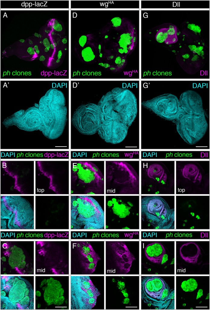
Fig 6. Loss of PRC1 is not sufficient for generalized de-repression of PcG target genes.
Expression of three known PcG targets was examined in discs carrying ph mutant clones, namely dpp (left panels), wingless (central panels) and Distal-less (right panels). (A-A’) Expression of the dpp-lacZ reporter (magenta) was restricted to stereotypical regions of wild-type discs near the morphogenetic furrow and a section of the antenna, despite the broad distribution of ph clones (GFP, green) in the disc. DAPI staining is shown in cyan. (B-C) Close-up sections of the same eye disc show that the dpp-lacZ reporter (magenta) was observed in sections above the GFP-marked ph clone (green) (top) (B), but in a middle section (C) where the cells lacking ph are visible (green) the expression of dpp-lacZ is interrupted and only detected in the surrounding wild-type cells. (D-D’) Staining for endogenously HA-tagged Wg (magenta) was not observed in the majority of ph clones (green). (E-F) close-up sections of eye discs show wgHA signal in a characteristic posterior region of the eye where photoreceptors normally differentiate, and reduced signal is detected in ph mutant cells arising in close proximity or within these regions. (G-G’) Dll staining (magenta) is restricted to the antenna region even in eye discs with ph clones (green), which did not show ectopic Dll signal. (H,I) The stereotypical Dll signal in the antenna is clear in a top section of a disc (H), but closer to the middle of the same disc (I) the growth of ph clones (green) in this region led to interruption of Dll staining in ph mutant cells. Scale bars represent 200 μm.

