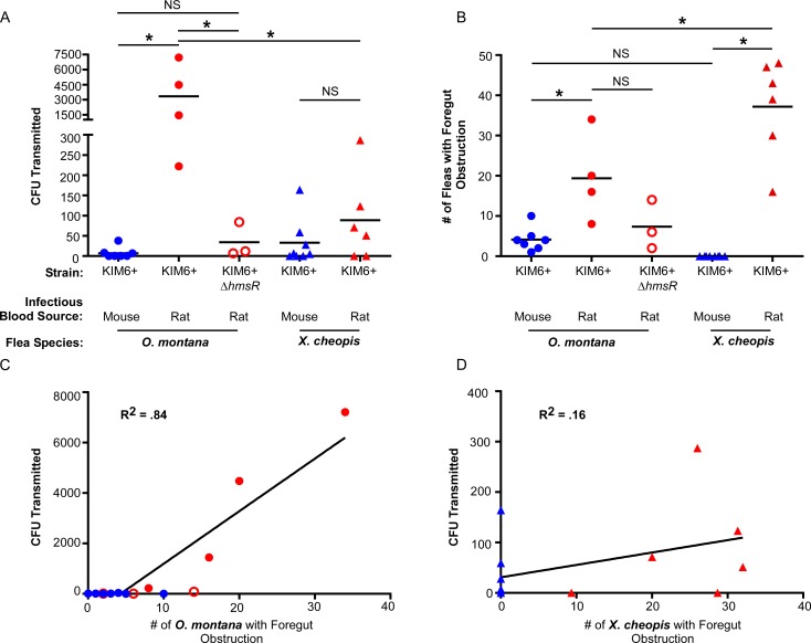
Fig 6. Biofilm-dependent transmission can augment early-phase transmission, but is dependent on flea species and infectious blood meal source.
(A) Y. pestis CFU transmitted in in vitro mass transmission experiments by groups of O. montana or X. cheopis 3 days after infection with Y. pestis KIM6+ (pAcGFP1) or KIM6+ ΔhmsR pAcGFP1) in mouse (blue symbols) or rat blood (red symbols). (B) Number of infected fleas from each transmission experiment that were diagnosed as either partially or completely blocked after feeding (fresh red blood present in both the esophagus and midgut or in the esophagus only). (C) Linear regression of the number of infected fleas with foregut obstruction (partial or complete blockage) and CFU recovered following transmission assays for O. montana (p < 0.0001) and (D) X. cheopis (p = 0.15). The coefficient of determination (R2) is listed on the graph. Each symbol on all four graphs represents data from a single independent transmission test (see Table 2 for details). Horizontal bars (A and B) indicate the mean; *p < 0.01 (A) or *p < 0.05 (B) by one-way ANOVA with Tukey’s post-test. NS = not significant. CFU data for mouse blood experiments in (A) are previously published [6].

