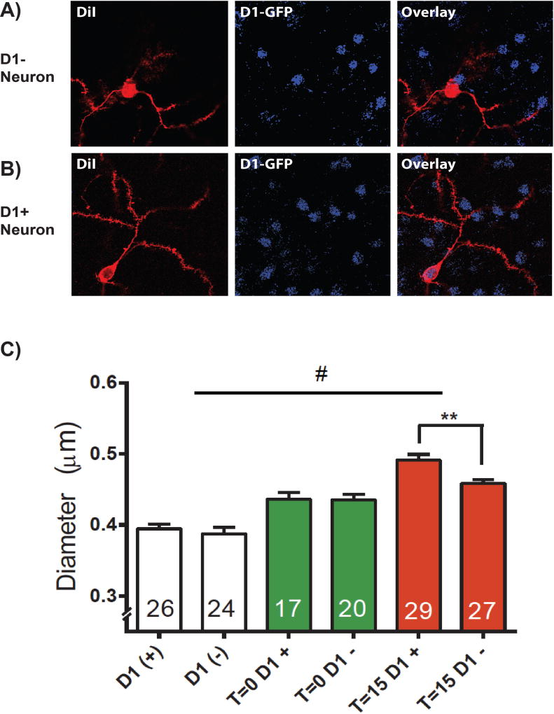
Figure 4.
DiI labeling coupled with Immunohistochemistry enhanced labeling of GFP allowed visualization of A) putative D2 neurons (D1 negative, D1−) and B) D1 positive neurons in the NAc C) Spine head diameter on D1+ and D1− neurons. After extinction from cocaine self administration (T=0, green bars), a potentiation in dh is observed in both D1+ and D1− neurons compared to saline controls (white bars). During cue-induced reinstatement (T=15, red bars), dh is elevated specifically on D1+ dendritic spines. N shown in bars is the number of neurons quantified, and the data were analyzed using a 2-way ANOVA F(1,137) = 4.613, p < 0.05 (main effect); ** D1 vs D2 p<0.01; # Between groups p< 0.001)

