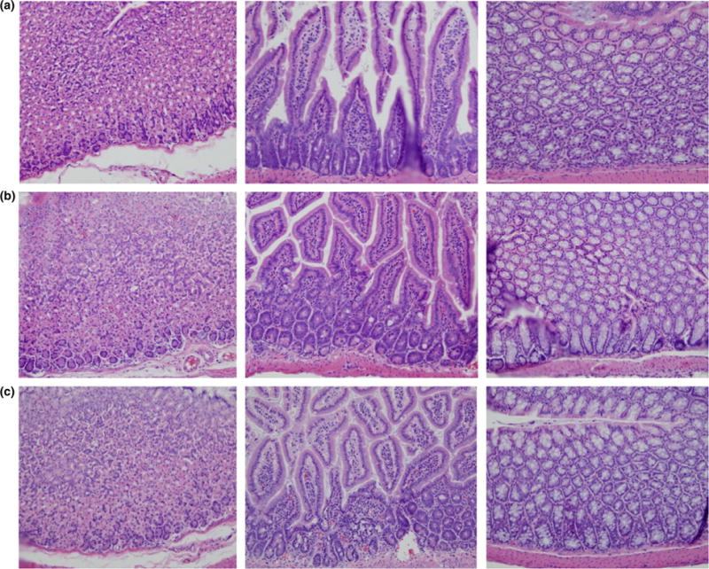
Figure 5.

Histological examination of tissues from mice challenged with Sulfolobus monocaudavirus 1 (SMV1) and M13KE. Histopathological observation of tissues obtained from stomach, small intestine and colon, demonstrating no treatment-related microscopic effects. (a). Tris-acetate control; (b). Bacteriophage M13KE; (c). Archaeal virus SMV1. [Colour figure can be viewed at wileyonlinelibrary.com]
