Figure 2.

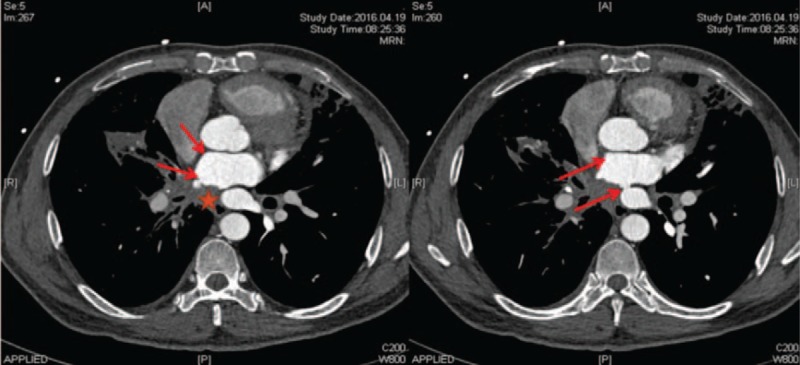
Chest CT (mediastinal window) in patient with FM. Multiple soft-tissue shadows (asterisk) are evident. Left and right superior PV occlusion and inferior PV stenosis were observed (arrows).

Chest CT (mediastinal window) in patient with FM. Multiple soft-tissue shadows (asterisk) are evident. Left and right superior PV occlusion and inferior PV stenosis were observed (arrows).