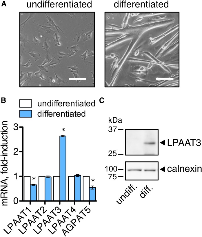
Fig. 1.
LPAAT3 was upregulated during differentiation of the C2C12 myoblast cell line. C2C12 cells were differentiated in low serum medium for 7 days, and then the myotubes were collected and differentiated for an additional 2 days. Undifferentiated cells were cultured in growth medium. A: Images of undifferentiated and differentiated C2C12 cells are shown. Scale bar = 100 μm. B: LPAAT3 mRNA was increased during differentiation. Expression levels were measured by RT-PCR. Data are expressed as the mean ± SEM from three independent experiments. *P < 0.05; t-test. C: LPAAT3 protein levels increased during differentiation. LPAAT3 was detected by Western blot analyses. Calnexin was detected as a loading control. Representative blots of three independent experiments are shown.

