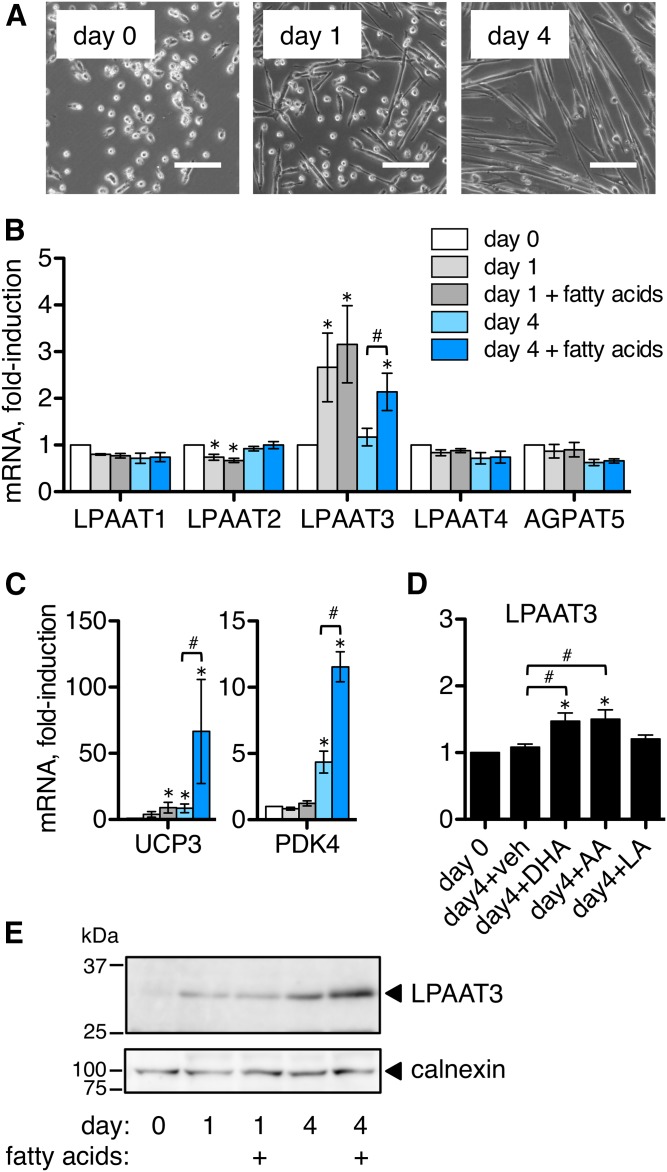
Fig. 2.
LPAAT3 levels were upregulated during satellite cell differentiation, and expression was enhanced by fatty acid supplementation. Satellite cells were differentiated in low serum medium, and some media were supplemented with a fatty acid mixture of LA, AA, and DHA (5 μM each). mRNA expression levels were measured by RT-PCR and protein levels were measured by Western blot. A: Images of satellite cells during differentiation. Satellite cells elongated and began fusing by day 1 and formed multinucleated myotubes by day 4. Scale bar = 100 μm. B–D: mRNA expression levels during satellite cell differentiation. B: LPAAT3 mRNA peaked at day 1 and expression was enhanced at day 4 by fatty acid supplementation. C: PPAR target genes UCP3 and PDK4 were also upregulated by fatty acid supplementation, suggesting possible PPAR activation. D: Components of the fatty acid mixture were applied individually (5 μM) during 4 days of differentiation. Either DHA or AA enhanced LPAAT3 expression. E: LPAAT3 protein increased during satellite cell differentiation and levels were enhanced by fatty acid supplementation at day 4. Representative Western blot from three independent experiments is shown; calnexin was detected as a loading control. RT-PCR data represent the mean ± SEM from three or four independent experiments. *P < 0.05, differentiated versus undifferentiated; #P < 0.05, fatty acid supplementation versus no supplementation; one–way ANOVA.

