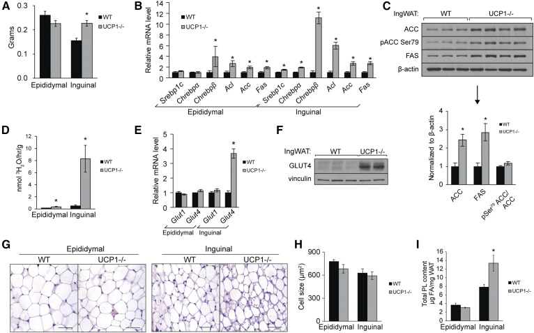
Fig. 2.
Ucp1 deletion causes inguinal WAT expansion and DNL. Twelve-week old chow-fed male mice were fasted 4 h prior to euthanasia. A: Weights of epididymal and inguinal WAT of UCP1−/− mice (n = 8–12/group). B: Expression of genes involved in FA synthesis in WAT (n = 6/group). C: Immunoblotting of lipogenic enzymes in inguinal WAT. D: In vivo rates of DNL in white adipose tissue depots. Twelve-week-old old male mice were fasted 2 h and 50 mCi of 3H2O was injected intraperitoneally. Mice were euthanized 2 h after injection (n = 3–7/group). E: Gene expression of glucose transporters in WAT depots (n = 6/group). F: Immunoblotting of GLUT4 in inguinal WAT of WT and UCP1−/− mice. G: WAT sections from WT and UCP1−/− were paraffin-embedded, sectioned, stained with H&E, and imaged with 40× magnification. Scale bar represents 50 µm. H: Average adipocyte size in WAT depots (n = 3/group) as measured by Adiposoft (33). I: Total PL content per milligram WAT as measured by TLC-GC (n = 6/group). Data are expressed as mean ± SEM. *P < 0.05 vs. WT.

