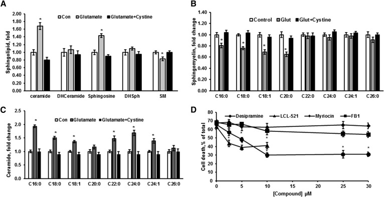
Fig. 4.
Sphingolipid changes in glutamate-treated OLs indicate activation of SM hydrolysis. Sphingolipids were analyzed in OLs treated with 1 mM glutamate alone and in the presence of 1 mM cystine for 24 h. Total ceramide, sphingosine, SM, dihydroceramide (DHCeramide), and dihydrosphingosine (DHSph) (A), SM species (B), and ceramide species (C) were quantified. Data are mean ± SE, *P < 0.05, n = 8. Each sample was normalized to its respective total protein levels. D: OLs were exposed to 1 mM glutamate with/without inhibitors of de novo sphingolipid synthesis, including myriocin and FB1 or SM hydrolysis pathway (desipramine and LCL-521) for 24 h and relative cell survival was determined. Data are mean ± SE, *P < 0.05, n = 12.

