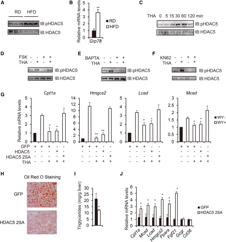
Fig. 4.
Obesity-induced ER stress suppresses fatty acid oxidation through phosphorylation of HDAC5. A: Immunoblot analysis of hepatic phosphorylated HDAC5 levels in RD-fed and HFD-fed mice under fasted state. B: Real-time PCR analysis of hepatic Grp78 mRNA amounts in RD-fed and HFD-fed mice (n = 3). C: Immunoblot showing effects of THA (500 nM) treatment on HDAC5 phosphorylation in primary hepatocytes at indicated times. D: Immunoblot showing effect of THA (500 nM, 1 h) treatment on FSK (10 μM)–induced HDAC5 dephosphorylation in primary hepatocytes. E: Immunoblot showing effect of BAPTA (1,2-bis(2-aminophenoxy)ethane-N,N,N′,N′-tetraacetate, 10 μM, 1 h) treatment on THA-induced HDAC5 phosphorylation in primary hepatocytes. F: Immunoblot showing effect of KN62 (10 μM, 1 h) treatment on THA-induced HDAC5 phosphorylation in primary hepatocytes. G: Effect of THA on mRNA amounts for WY-induced fatty acid oxidation genes, including Cpt1a, Hmgcs2, Lcad, and Mcad in primary hepatocytes reconstituted with HDAC5 or HDAC5 2SA (n = 3). H: Representative Oil Red O staining of liver sections from HFD-fed mice injected with either Ad-GFP or Ad-HDAC5 2SA (scale bar, 20 μm). I: Effect of Ad-GFP or Ad-HDAC5 2SA injection on hepatic triglyceride levels in HFD-fed mice (n = 8). J: Effect of Ad-GFP or Ad-HDAC5 2SA injection on hepatic fatty acid oxidation gene expression in HFD-fed mice (n = 8). All data are presented as means ± SEM. *P < 0.05. **P < 0.01.

