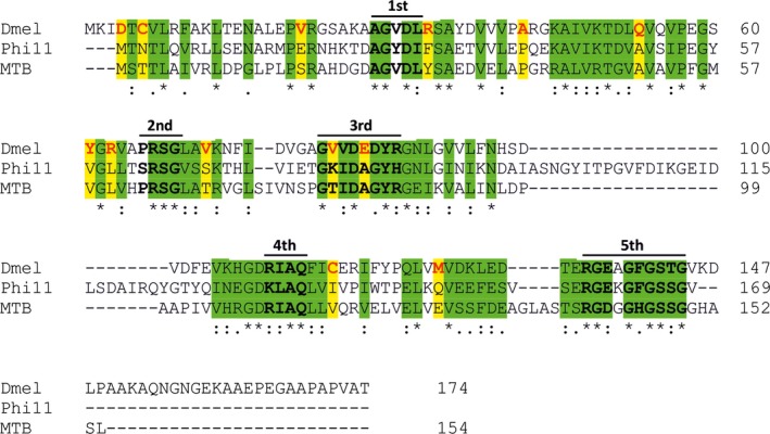
Figure 5.

Species‐specific sequence similarities and alterations among three Stl‐inhibited all‐β dUTPases. Conserved sequence motifs are highlighted with bold black case and upper lines. Amino acids with similar side‐chain characteristics are shown in green background. Residues that are similar to Φ11 and M. tuberculosis dUTPases but different in the Drosophila melanogaster enzyme have yellow background. Drosophila melanogaster dUTPase residues with alternate characteristics are emphasized with red letters.
